Instruments
BD FACSAria-SORP
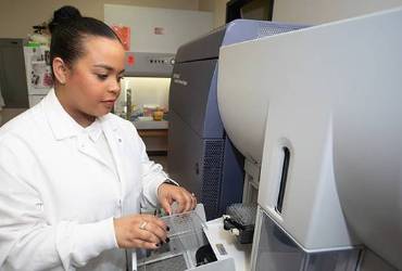 The FACSAria-SORP cell sorters are capable of 12 fluorescence parameters. The sorters
have 4 laser lines, including a 60 mW 350 nm UV line, 100 mW 488 nm, 100 mW 561 nm,
and 40 mW 640 nm lines. The sorters can be used to isolate cells in multiple formats,
including 4-way tube sorting and 96-well plate sorting via the Automated Cell Deposition
Unit.
BD FACSCanto II
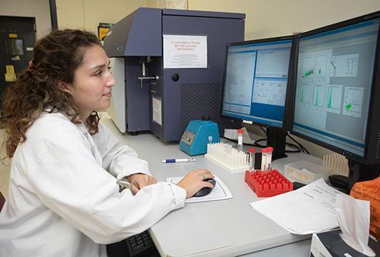 The FACSCanto II cell analyzers are equipped with 3 lasers (30 mW 405 nm, 20 mW 488
nm, and 17 mW 640 nm) capable of 8 fluorescence parameters. The analyzer is available
to individual investigators. Appropriate dichroics are available for numerous routine
as well as specialized assays for each of the instruments.
Nexcelom Celigo microplate based imaging cytometer
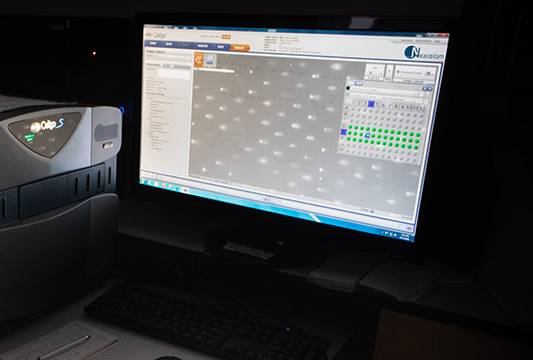
The CeligoS imaging cytometer is designed for performing assays directly in multi-well plates (6-384 wells). There are many advantages to this approach, including lower demand for cells (allowing for the use of rare primary cells isolated from patient samples) and reagents as well as analyzing the cells in their native state without the need to enzymatically remove them from their growth substrate, as is required for traditional flow cytometry. It can image cells in Brightfield and 3 fluorescent channels: blue (377/50nm Ex 470/22nm Em), green (483/32 Ex 536/40 Em), and orange/red (531/40 Ex 629/53 Em). Using advanced software based image recognition, there are over 10 specific analysis modes for a wide variety of applications. The CeligoS is also a self-serve instrument after training is provided.
XRAD320
https://www.kimtron.com/
On-Chip Biotechnologies Microfluidic Cell Sorter
(3 laser 6 color)
The On-Chip is the world’s first in its class microfluidic cell sorter and was developed in Japan. USA MCI was one of the first centers in America to obtain this new technology. One benefit over traditional sorting is the ability to analyze extremely low starting sample volumes/number of cells. Additionally, the sorting is much gentler and can accommodate a wide range of particles from exosomes to cell spheres using two different size microfluidic chips. The sorter is aerosol free and placed in a BSL-2 cabinet, allowing for the safe isolation of cells infected with lenti virus or other pathogens. The 3 excitation lasers (405nm, 488nm & 561nm) are collinear, so fluorochromes excited by any laser can be detected in any of these channels: FL1(445/20 nm), FL2(543/22 nm), FL3(591.5/43 nm), FL4(607/36 nm), FL5(676/37 nm), and FL6(732/68 nm).
On-Chip Biotechnology SPiS
The SPiS companion instrument does single cell or sphere deposition into 96 or 384 well plates in a far more gentle and reliable manner than traditional sorters.
ThermoFisher Neon Electroporation System
Designed for primary cells and large size plasmids, very gentle. Available for use free of charge with appointment.
Miltenyi Biotech GentleMACS tissue disassociation system
The industry standard for preparing single cell suspensions from most any tissue with the highest viability. Available for use free of charge with appointment.
Agilent Quanteon Flow Cytometer
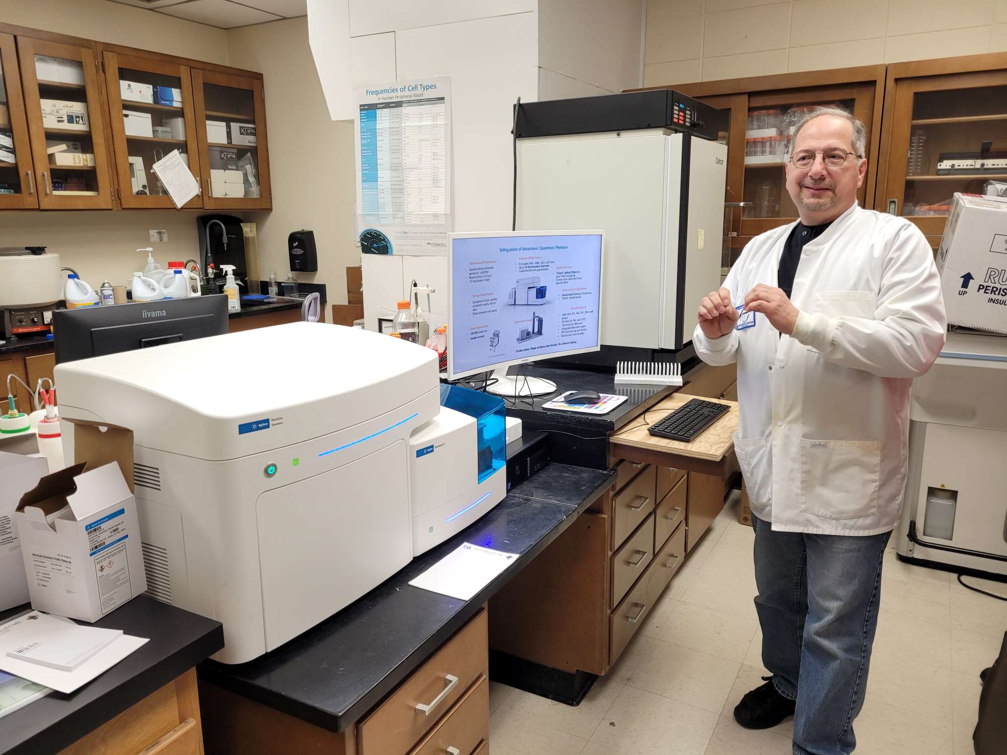
detection. It uses SiPM detectors making it one of the most sensitive instruments available. This sensitivity enables the accurate detection of very small particles such as viruses and exosomes in the 100nm size range. The Quanteon is equipped with an advanced sample autoloader, able to sample from a rack of 40ml tubes, or 96 well plates (regular and deep-well) and 384 well plates. Usage rates are $25/hour unassisted, $40/hour assisted within the university and $75/hour for ‘outside’ users.
The ZetaView® TWIN - NTA Nanoparticle Tracking - Video Microscope PMX-220
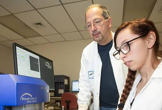
Tracking Analysis (NTA) captures the Brownian motion of each particle in the video. Based on
the different diffusion movements of large and small particles in the surrounding liquid, the hydrodynamic diameter of the particles is determined. Pattern parameters, such as intensity
fluctuations, surface geometry and shape of the particles as well as particle concentration are
documented at each recording and can be used to distinguish sub-populations. In addition, the
charge state of the particle surface (zeta potential) can be measured via the movement of the particles in an applied electric field. Depending on the type of sample and the measuring mode, the measuring range is between 15 nm and 5 μm. User rates are $25/hour.
The Agilent Seahorse XFe24 Analyzer
Kimtron IC-320

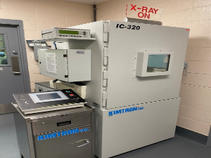
X-ray cabinet irradiation systems are designed to deliver precise X-ray dosages to specimens that range in size from cells to small animals. Both COM SRL locations have nearly identical irradiators (Kimtron IC-320 at MSB; XRAD320 at MCI). The systems are safe but require assistance and training before independent operation is permitted. Applications are varied and include production of non-dividing feeder cells, X-ray virus inactivation, production of sterile insects for reducing insect populations, and X-ray sterilization of items that cannot be steam sterilized.
10x Genomics Chromium Controller
Miltenyi Biotech GentleMACS tissue disassociation system
The industry standard for preparing single cell suspensions from most any tissue with the highest viability. Available for use free of charge with appointment.
Finesse 325 Microtome
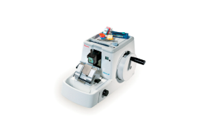
Formalin-fixed paraffin embedded (FFPE) tissues can be sectioned using the Finesse 325 microtome (Thermo Scientific) located in the MSB facility. FFPE sections can be examined using various histological and immunohistological approaches.
Crystostat
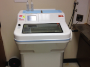
A cryostat (Thermo Scientific) is available for sectioning frozen tissues. Frozen sections can be examined by various histological and immunohistological methods.


